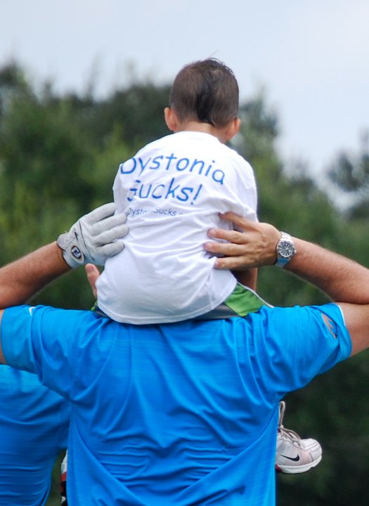
About Dystonia
Dystonia is a neurological movement disorder characterized by involuntary muscle contractions, which force certain parts of the body into abnormal, sometimes painful, movements or postures. Dystonia can affect any part of the body including the arms and legs, trunk, neck, eyelids, face, or vocal cords. Abilities such as cognition, strength and the senses are normal in Dystonia sufferers, though speech can be impaired as a symptom. Dystonia is not fatal, but is a chronic disorder with often unpredictable prognoses. Dystonia is the third most common movement disorder after Parkinson’s Disease and Tremor. Dystonia does not discriminate: it affects people of every race and ethnic group and one-third of Dystonia patients are children. Dystonia affects more people than Muscular Dystrophy, Huntington’s Disease and Lou Gehrig’s Disease combined.
Dystonia News
Long-Term Outcomes of Deep Brain Stimulation for Pediatric Dystonia
Background: Deep brain stimulation (DBS) has been utilized for over two decades to treat medication-refractory dystonia in children. Short-term benefit has been demonstrated for inherited, isolated, and idiopathic cases, with less efficacy in heredodegenerative and acquired dystonia. The ongoing publication of long-term outcomes warrants a critical assessment of available information as pediatric patients are expected to live most of their lives with these implants. Summary: We performed a review of the literature for data describing motor and neuropsychiatric outcomes, in addition to complications, 5 or more years after DBS placement in patients undergoing DBS surgery for dystonia at an age younger than 21. We identified 20 articles including individual data on long-term motor outcomes after DBS for a total of 78 patients. In addition, we found five articles reporting long-term outcomes after DBS in 9 patients with status dystonicus. Most patients were implanted within the globus pallidus internus, with only a few cases targeting the subthalamic nucleus and ventrolateral posterior nucleus of the thalamus. The average follow-up was 8.5 years, with a range of up to 22 years. Long-term outcomes showed a sustained motor benefit, with median Burke-Fahn-Marsden dystonia rating score improvement ranging from 2.5% to 93.2% in different dystonia subtypes. Patients with inherited, isolated, and idiopathic dystonias had greater improvement than those with heredodegenerative and acquired dystonias. Sustained improvements in quality of life were also reported, without the development of significant cognitive or psychiatric comorbidities. Late adverse events tended to be hardware-related, with minimal stimulation-induced effects. Key Messages: While data regarding long-term outcomes is somewhat limited, particularly with regards to neuropsychiatric outcomes and adverse events, improvement in motor outcomes appears to be preserved more than 5 years after DBS placement.
TorsinB overexpression prevents abnormal
twisting in DYT1 dystonia mouse models
twisting in DYT1 dystonia mouse models
Genetic redundancy can be exploited to identify therapeutic targets for inherited disorders. We explored this possibility in DYT1 dystonia, a neurodevelopmental movement disorder caused by a loss-of-function (LOF) mutation in the TOR1A gene encoding torsinA. Prior work demonstrates that torsinA and its paralog torsinB have conserved functions at the nuclear envelope. This work established that low neuronal levels of torsinB dictate the neuronal selective phenotype of nuclear membrane budding. Here, we examined whether torsinB expression levels impact the onset or severity of abnormal movements or neuropathological features in DYT1 mouse models. We demonstrate that torsinB levels bidirectionally regulate these phenotypes. Reducing torsinB levels causes a dose-dependent worsening whereas torsinB overexpression rescues torsinA LOF-mediated abnormal movements and neurodegeneration. These findings identify torsinB as a potent modifier of torsinA LOF phenotypes and suggest that augmentation of torsinB expression may retard or prevent symptom development in DYT1 dystonia.
Research at DukeDuke researchers develop new cell-based drug screening test for dystoniaPublished on December 8, 2016 Duke University researchers have identified a common mechanism underlying separate forms of dystonia, a family of brain disorders that cause involuntary, debilitating and often painful movements, including twists and turns of different parts of the body.
Dystonia is the third most common movement disorder, after Parkinson's disease and tremors. It is believed to affect 300 to 400 people per million population, and is more common in the elderly. The spectrum of dystonia's many distinct forms include rare inherited cases like DYT1 dystonia, which is caused by a specific mutation in a single gene and leads to a severe disorder that arises in childhood. A larger proportion of cases, grouped into non-familial dystonia, have no known cause and tend to occur during adulthood. In the new study, the team uncovered a mechanism for dystonia that links together rare inherited dystonia with the more common non-familial cases. To learn more, please click here. Novel Therapy Development for Dystonia
Dystonia is a movement disorder characterized by involuntary repetitive, sustained muscle contractions, or postures. About 300,000 to 500,000 individuals, including military and veteran populations, suffer from dystonia in the US. DYT1 dystonia is the most common type among the genetic dystonia as an early-onset hyperkinetic movement abnormality. This type of dystonia is prevalent in ages from 5 to 28 years old. The symptoms of DYT1 dystonia start from limbs and then remain segmental or become generalized with cervical muscle involvement. Patients usually are severely disabled and limit to wheelchairs. The DYT1 dystonia is an autosomal dominant disease showing variability in phenotype with low penetrance (30%-40%) possibly caused by additional genetic mutations and/or environmental variations. The individuals affected by DYT1 dystonia share the same
genetic mutation located in exon 5 of DYT1 gene, leading to a loss of an amino acid residue for a protein called torsinA. This type of mutation occurs in 50-60% of non-Jewish patients and more than 90% of Ashkenazi Jewish patients. DYT6 is another form of generalized, early-onset dystonia. Unlike DYT1 dystonia, DYT6 dystonia symptoms appear with a slower progression and frequently involve the cervical and cranial muscles. In addition, DYT6 does not respond well to deep brain stimulation (DBS) in the brain. In DYT6, the causative gene is THAP domain-containing apoptosis-associated protein 1 (THAP1). The THAP1 is a small DNA binding protein. There are commonalities between DYT1 dystonia and DYT6 dystonia. Thap1 appears to regulate the activities of torsinA. Furthermore, brain imaging studies show that a certain connection in the brain influences penetrance in both DYT1 and DYT6 dystonia. The current treatments for dystonia have variable success rates among individuals and even in the same person over time. We aim to develop and validate a novel virus-based treatment strategy based on the role of this brain connection in influencing the penetrance and dystonia severity. We plan to use an overt dystonia mouse model (Dlx-CKO mice) to test the hypothesis that Purkinje cell-specific viral expression of DREADD (designer receptors exclusively activated by designer drugs) molecules will alter this connection and improve motor performance and dystonia in an overt dystonia mouse models. It should be noted that although we are using the Dlx-CKO mice that were developed using conditional knockout of torsinA, this overt dystonia mouse model should have a broad representation of other types of dystonias as well because knockout of torsinA in Dlx5/6-positive neurons does not occur in humans. Cerebellar stimulation has been used in treating dystonia patients in the past with mixed success. The advantage of using the DREADD approach is that it not only can stimulate neuronal activity; it can also silence the neuronal activity. Furthermore, it can manipulate specific types of neurons or over multiple brain regions or anatomic locations, all depends on the specific gene instructions used that are available to the scientists. Viral therapies recently reached a milestone when the FDA approved Novartis’ Zolgensma in May 2019 for the treatment of children with spinal muscular atrophy (SMA). The therapy approved is to introduce the missing gene that can help in the production of the protein that is otherwise missing in patients with SMA. As clinical efficacy of virus-based vectors becomes more established, there is an expectation that applications of these vectors will extend to other neurological indications, including dystonia. The successful completion of the proposed experiments will pave the way for the future development and implementation of virus-based treatment for DYT1, DYT6, and other dystonias. This may include testing in a larger animal model that is closer to human patients, applying for IND, and starting phase 1 trial in patients. |
Research at VIB in Belgium Prof. Rose Goodchild (VIB-KU Leuven): "For the first time, we understand that a dystonia protein is responsible for cellular lipid levels. Although we had expected a more complex picture, with various direct and indirect effects, our data clearly labeled torsin as the regulator for a particular enzyme of lipid metabolism. This now focuses attention on how the lipid substrates and products of this enzyme contribute to neuronal function, and gives us a better view on the exact molecular defects that cause dystonia."
To learn more, please click here. New Imaging Technique Could Aid in Testing of New Drugs for DystoniaA new study led by University of Florida neuroscientists furthers the scientific understanding about the brain regions involved with causing dystonia, a poorly understood, debilitating neurological disorder that causes involuntary muscle contractions, twisting movements and other symptoms. The new findings may help scientists test new drugs that could help abate the symptoms of the disease.
The study, published in the journal Neurobiology of Disease, shows that removing a key protein linked to dystonia in a mouse model results in widespread increases in connectivity strength across the brain network. The protein is called torsinA and is associated with DYT1 dystonia, which is a genetic form of dystonia. To learn more, please click here Mutant Allele-Specific CRISPR Disruption in DYT1 Dystonia Fibroblasts Restores Cell FunctionMost individuals affected with DYT1 dystonia have a heterozygous 3-bp deletion in the TOR1A gene (c.907_909delGAG). The mutation appears to act through a dominant-negative mechanism compromising normal torsinA function, and it is proposed that reducing mutant torsinA may normalize torsinA activity. In this study, we used an engineered Cas9 variant from Streptococcus pyogenes (SpCas9-VRQR) to target the mutation in the TOR1A gene in order to disrupt mutant torsinA in DYT1 patient fibroblasts. Selective targeting of the DYT1 allele was highly efficient with most common non-homologous end joining (NHEJ) edits, leading to a predicted premature stop codon with loss of the torsinA C terminus (delta 302–332 aa). Structural analysis predicted a functionally inactive status of this truncated torsinA due to the loss of residues associated with ATPase activity and binding to LULL1. Immunoblotting showed a reduction of the torsinA protein level in Cas9-edited DYT1 fibroblasts, and a functional assay using HSV infection indicated a phenotypic recovery toward that observed in control fibroblasts. These findings suggest that the selective disruption of the mutant TOR1A allele using CRISPR-Cas9 inactivates mutant torsinA, allowing the remaining wild-type torsinA to exert normal function.
|

